WATERS OF LIFE
LIFE UNDERWATER
- VEGETATION
- PLANKTON
- BENTHOS
- INTRODUCTION - BENTHOS
- BIODIVERSITY
- ENVIRONMENT AND ADAPTATIONS
- NUTRITION
- DISTRIBUTION
- RESEARCHER'S OPINION
- FISH
- AMPHIBIANS AND REPTILES
BENTHOS - ENVIRONMENT AND ADAPTATIONS
![]() FLASH PLAYER IS REQUIRED TO SEE THE CONTENT OF THIS PAGE
FLASH PLAYER IS REQUIRED TO SEE THE CONTENT OF THIS PAGE
VIDEO - 0 min 11 s
This small, whitish leech is a parasite on this gastropod.
This page contains videos that require Javascript and Adobe Flash Player to be activated. If you wish not to activate these functions, we offer pictures from the original videos.

VIDEO - 0 min 12 s
The protozoans covering this isopod’s body are parasitising it.
This page contains videos that require Javascript and Adobe Flash Player to be activated. If you wish not to activate these functions, we offer pictures from the original videos.

VIDEO - 0 min 10 s
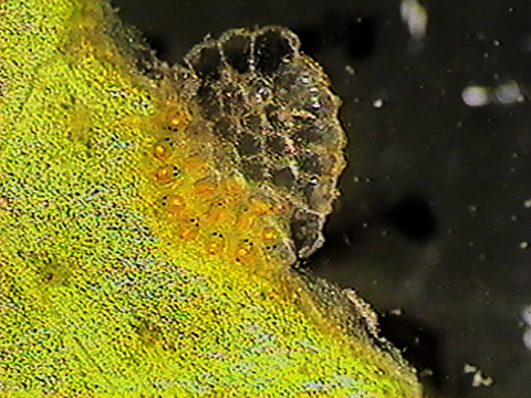
Water mites deposit their eggs in gelatinous masses on plants in habitats near potential prey for the young to parasitise when they hatch.
This page contains videos that require Javascript and Adobe Flash Player to be activated. If you wish not to activate these functions, we offer pictures from the original videos.

VIDEO - 0 min 22 s
Amphipods rapidly move their legs to create a current and bring more oxygen to their gills.
This page contains videos that require Javascript and Adobe Flash Player to be activated. If you wish not to activate these functions, we offer pictures from the original videos.

VIDEO - 0 min 20 s
Planarians (flatworms) have no gills, and breathe through osmosis. Water is absorbed into the planarian’s body through its skin. Planarians can also regenerate lost body parts. Planarians are hermaphroditic; each individual is both male and female.
This page contains videos that require Javascript and Adobe Flash Player to be activated. If you wish not to activate these functions, we offer pictures from the original videos.

VIDEO - 0 min 10 s
To facilitate gas transfer between their skin and the water, the worm undulates its entire body.
This page contains videos that require Javascript and Adobe Flash Player to be activated. If you wish not to activate these functions, we offer pictures from the original videos.

VIDEO - 0 min 08 s
By contorting their bodies, caddisfly larvae increase water circulation through their case, boosting the amount of oxygen that arrives to their gills.
This page contains videos that require Javascript and Adobe Flash Player to be activated. If you wish not to activate these functions, we offer pictures from the original videos.

VIDEO - 0 min 07 s
Because deep sediments have very low oxygen concentrations, some organisms, including chironomids (blood worms), have had to evolve the ability to make hemoglobin.
This protein transports oxygen within the organism, increasing its ability to absorb oxygen and giving it a reddish colour.
WWW | USEFUL LINKS - WATER MONITORING YOUTH PORTAL
This page contains videos that require Javascript and Adobe Flash Player to be activated. If you wish not to activate these functions, we offer pictures from the original videos.

VIDEO - 0 min 13 s
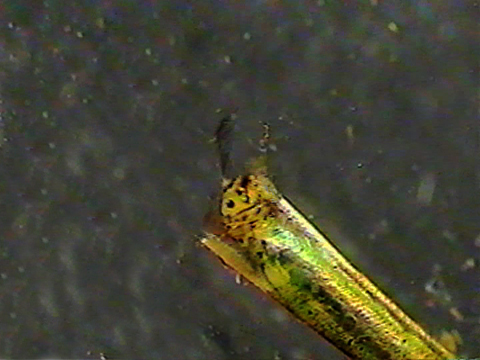
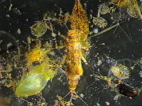
This mayfly larva’s gills are located on the dorsal part of its abdomen and are protected by two opercula.
This page contains videos that require Javascript and Adobe Flash Player to be activated. If you wish not to activate these functions, we offer pictures from the original videos.

VIDEO - 0 min 07 s
Diving beetle larvae rise to the surface and use the respiratory siphons (urogomphi) on the tip of their abdomen to collect air. This air is kept in reserve, like the oxygen tank of a scuba diver.
This page contains videos that require Javascript and Adobe Flash Player to be activated. If you wish not to activate these functions, we offer pictures from the original videos.

VIDEO - 0 min 10 s
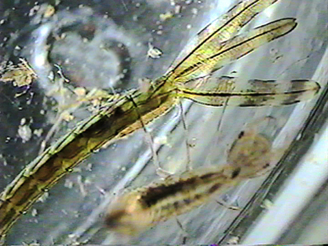
Zygopteran (damselfly) larvae breathe through three gill lamellae located at the tip of the abdomen.
This page contains videos that require Javascript and Adobe Flash Player to be activated. If you wish not to activate these functions, we offer pictures from the original videos.

VIDEO - 0 min 17 s
Anisopterans (dragonfly) larvae have a muscular rectal chamber which they can fill with water. This rectal pump mainly serves for respiration, but can also be used for locomotion: the larva can contract its muscles, expelling the water and propelling the animal forward.
LIFE UNDERWATER
BENTHOS
- INTRO BENTHOS
- BIODIVERSITY
- ENVIRONMENT AND ADAPTATIONS
- NUTRITION
- DISTRIBUTION
- RESEARCHER'S OPINION
